Low-dose radiographs could reveal typical lung changes
New x-ray method for Corona diagnosis ready for patient testing
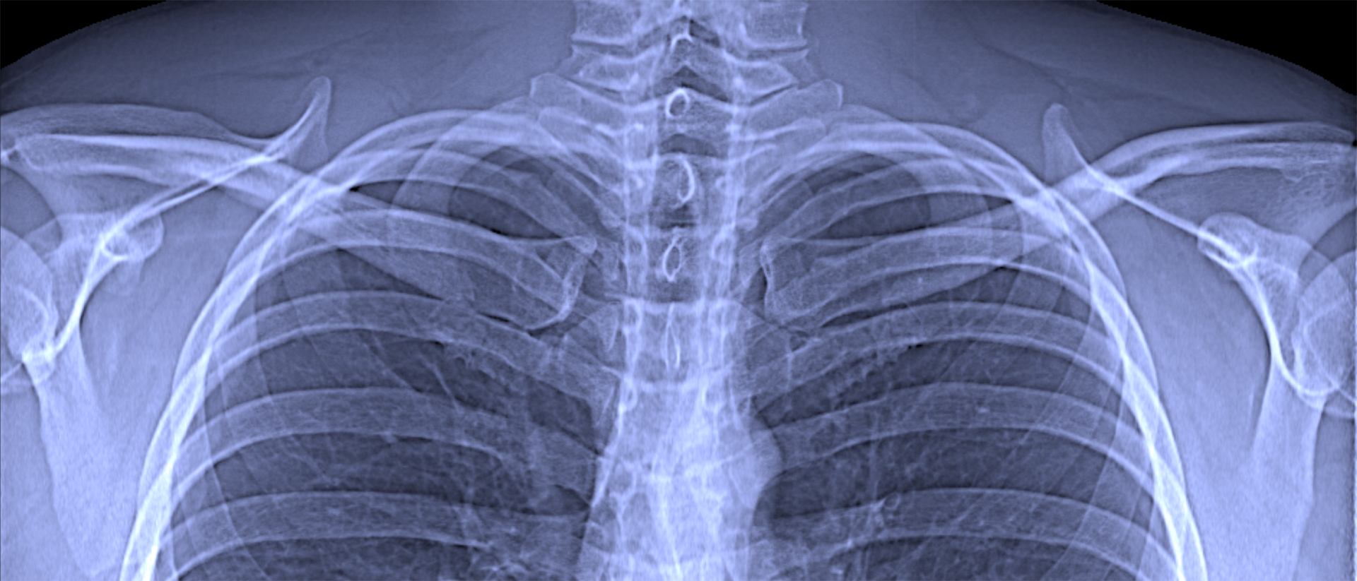
Reliable methods for identifying the new Coronavirus are crucial during the pandemic it has caused. In addition to biochemical tests, x-ray methods can also be used to identify pathological changes in the lung that can typically accompany Covid-19. These x-ray methods make it possible to examine large numbers of patients within a very short time, providing results immediately after the examination.
Deflected x-rays exposes areas with damaged pulmonary alveoli
Working together with colleagues from the university hospital TUM Klinikum rechts der Isar, Franz Pfeiffer, Professor for Biomedical Physics and Director of TUM’s Munich School of BioEngnieering, now plans to test the new dark-field x-ray imaging procedure in the diagnosis of Covid-19.
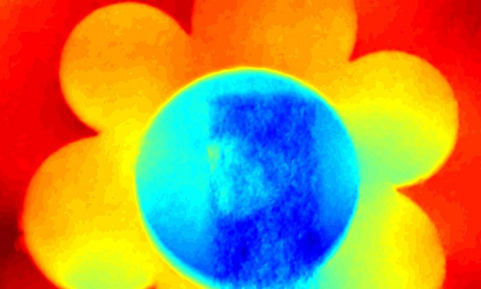
While conventional x-ray imaging shows the attenuation of x-rays on their way through tissue, the dark-field method focuses on the small share of the x-ray light which is scattered, i.e. diverted from its straight path. Conventional x-ray images ignore this scattered x-ray light.
The new method thus uses the physical phenomenon of scattering in a way similar to long-established dark-field microscopy technologies using visible light. They make it possible to clearly visualize objects which are for the most part transparent and which appear in the darkfield microscope as clear structures in front of a dark background, giving dark-field microscopy its name.
“The scattering is particularly strong for example at interfaces between air and tissue,” says Pfeiffer. As a result, a dark-field image of the lung can clearly distinguish areas with alveoli that are intact, i.e. filled with air, from regions in which the alveoli have collapsed or are filled with fluid.
In pneumonia of the type caused by Covid-19, structures form in the lung which initially resemble cotton wadding or spider webs and which then spread throughout the lung and fill with fluid. In combination with other typical symptoms these structures are a clear indication of a Covid-19 infection. The changes in the lung are associated with damage to the alveoli which could be clearly visible in dark-field images.
Innovative method
Dark-field imaging with x-rays is a completely innovative examination method in medicine. Over a period of more than ten years Franz Pfeiffer and his team developed the method from its very beginnings. Professor Pfeiffer presented the fundamental approach, which makes it possible to use the method with conventional x-ray tubes of the type found in physician's offices, in 2008.
Until then the method required x-rays of a higher quality available only from synchrotron light sources – complex large research facilities. After the initial laboratory experiments, Pfeiffer and his staff members worked in close collaboration with physicians to develop the method further. Now a device, which is suitable for examining patients is available.
Low radiation doses
Examinations using dark-field technology would involve exposure to a significantly lower radiation dose than the computed tomography used today. This is because the new technology requires only a single image per patient, while computed tomography requires a large number of individual images taken from various angles. The changes caused by Covid-19 cannot be clearly identified in a conventional two-dimensional x-ray image.
Now that the Federal Office for Radiation Protection (Bundesamt für Strahlenschutz) has issued its approval, testing can begin within the next weeks. Here the researchers will offer patients at the university hospital TUM Klinikum rechts der Isar who are being examined for Covid-19 with computed tomography the chance to also undergo examination using the dark-field method.
This is intended to confirm that the illness can be reliably diagnosed using this approach. “We're very optimistic, since at present this method is also highly successful in tests for other illnesses which are associated with changes in the lung,” says Prof. Marcus R. Makowski, Director of the Institute for Diagnostic and Interventional Radiology at the university hospital TUM Klinikum rechts der Isar.
Accelerating development of equipment
Franz Pfeiffer hopes that these tests will speed up clinical studies and the development of marketable equipment that uses the dark-field method. “It would certainly be over a year before this kind of device was available. But we can assume that the demand for cost-effective, reliable and low-impact Covid-19 diagnostics will remain unchanged for quite some time,” Pfeiffer points out.
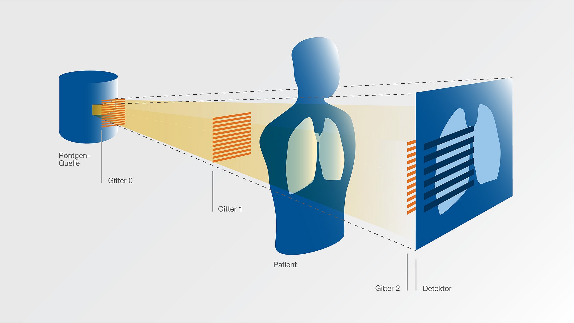
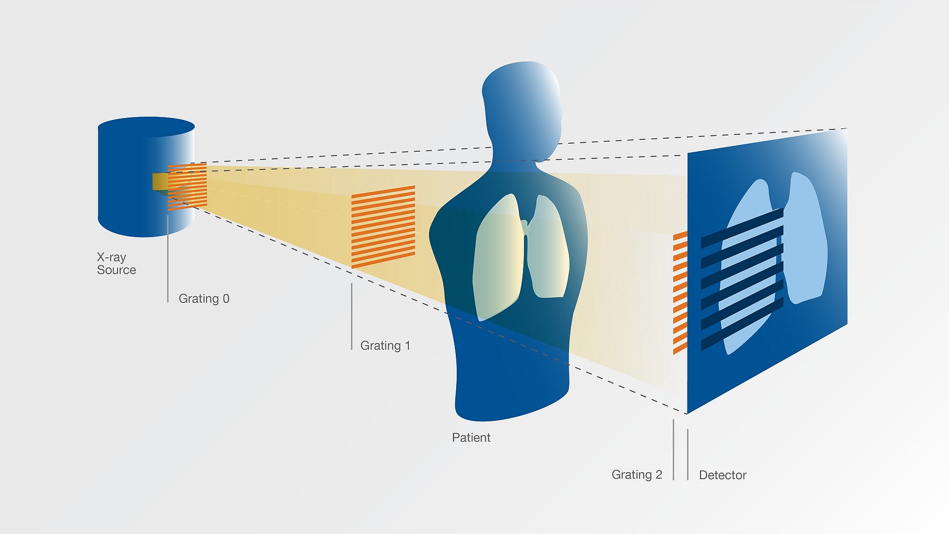
Description of grating-based x-ray dark-field imaging on the webpage of the Munich School of BioEngineering
High-resolution images
Technical University of Munich
Corporate Communications Center
- Paul Piwnicki
- paul.piwnicki@tum.de
- +49 89 289 10808
- presse@tum.de
- Teamwebsite
Contacts to this article:
Prof. Dr. Franz Pfeiffer
Technical University of Munich
Chair of Biomedical Physics
Munich School of BioEngineering
Phone: +49 171 301 9552
Email: franz.pfeiffer(at)tum.de

![[Translate to en:] Eine Kombination von Dunkelfeld- und konventionellem Röntgenbild ermöglicht eine klare Unterscheidung zwischen gesundem und emphysematösem Gewebe sowie eine Beurteilung der regionalen Verteilung der Krankheit. Anhand solcher Bilder k A combination of dark-field and conventional transmission information allows for a clear distinction of healthy versus emphysematous tissue and an assessment of the regional distribution of the disease. From such images, a doctor might in future not only see if a patient is diseased but also which parts of the lung are affected and how much. Picture: Simone Schleede / TUM](/fileadmin/_processed_/6/d/csm_121022_Dunkelfeld_900_28fbd8d6cb.jpg)
![[Translate to en:] Die Komponenten im Inneren des CT-Prototypen sind eine Röntgenquelle (links), ein Detektor (ganz rechts) und ein Gitterinterferometer für die Phasenkontrastbildgebung. Mounted in a movable gantry inside the CT scanner are an x-ray source (left), a detector (far right), and a three-grating interferometer for phase-contrast imaging. Image: A. Tapfer/TUM](/fileadmin/_processed_/7/2/csm_Vorschau_Photo_and_schematic_a3837ca3bd.jpg)