FRM II lets scientists look deep inside the structure of biomolecules
Neutrons for better vaccines against multidrug resistant germs
Bacteria which are resistant to all conventional antibiotics cause more than a million deaths each year. Consequently, researchers around the world are searching for new therapeutic approaches to combat these pathogens. Two years ago an international team in Grenoble identified an active ingredient suitable for the production of a vaccine against multidrug resistant bacteria Pseudomonas aeruginosa. The vaccine has in the meantime been successfully tested on mice.
"As with many new vaccines, in this case the active ingredient is embedded in liposomes. The exact characterization and understanding of these nanoscopic biomolecules is a key factor in the development and optimization of future vaccines," says Dr. Marco Maccarini, biophysicist at the French National Centre for Scientific Research (CNRS). Together with experts at the TIMC laboratory of the Université Grenoble Alpes (UGA) and at the FRM II he has successfully analyzed the structure of the candidate vaccine against Pseudomonas aeruginosa.
The vaccine consists of biomolecules with sizes in the order of 100 nanometers. These molecules consist mostly of lipids, substances similar to fats, which form small bubbles or liposomes because of their biochemical properties. These bubbles can in turn protect and transport the actual active ingredients. In the case of the vaccine against Pseudomonas aeruoginosa, this active ingredient is the protein OprF. "In general, the active ingredient can dock to the liposome at various positions – for example internally or externally," Maccarini explains. "But it is best recognized by the immune system when it is integrated in the double lipid layer. Thus the structure of the biomolecule is decisive in the efficacy of a vaccine."
Wanted: Non-destructive radiation
Such structural details are not visible to the naked eye. Optical microscopes do not have a high enough resolution for the investigation of liposomes. Although x-ray radiation has shorter wavelengths, it is unsuitable for structural analysis, since the radiation can damage the biomolecules under certain circumstances. "But neutron beams are ideal: They only interact with atomic nuclei and thus cause no damage or structural changes. This way the samples can be investigated in their original condition," Maccarini says.
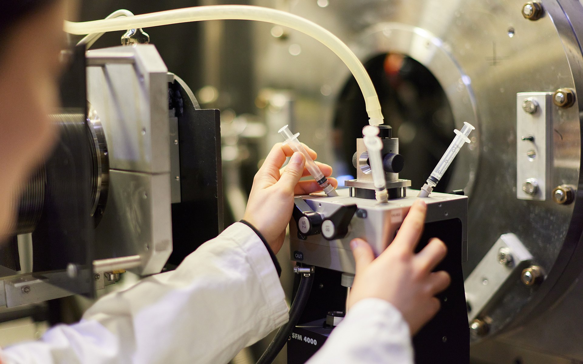
The researcher found everything he needed to analyze the new candidate vaccine at the FRM II in Garching near Munich: a high neutron flux, a well-equipped laboratory and Dr. Aurel Radulescu, an expert in the measurement of small-angle scattering, a technology which can be used to investigate the nanometer-sized molecules in detail.
Computer model represents structure of the vaccines
"In our case the challenge was using the diffractometer, which measures the scattering of the neutrons by the atomic nuclei, to distinguish between the proteins and the lipids in the sample," recalls Radulescu, who supervises the small-angle scattering diffractometer KWS-2 for the Forschungszentrum Jülich (FZJ) in Garching. He adds that it took a trick to finally make this distinction work: "We conducted the measurements with different combinations of solvents – normal water and heavy water containing deuterium, mixed in various concentrations." Since neutrons "see" normal hydrogen and deuterium differently, the result was images of the sample with different contrasts that contained distinct information.
For the analysis the research team developed a computer model which represents the structure of the candidate vaccine. "This lets us not only render the two-layer structure of the lipids, but also lets us determine the average position and amount of the OprF active ingredient which is embedded between the two lipid layers."
The new model can also be used to investigate the structure of new, liposome-based vaccines and to optimize their further development.
- Francesco Spinozzi, Jean-Pierre Alcaraz, Maria Grazia Ortore, Landry Gayet, Aurel Radulescu, Donald K. Martin, and Marco Maccarini: Small-Angle Neutron Scattering Reveals the Nanostructure of Liposomes with Embedded OprF Porins of Pseudomonas aeruginosa, Langmuir 2022, 38, 15026−15037
doi.org/10.1021/acs.langmuir.2c01342
- High-resolution pictures
- The research activities were supported by France's Investissements d'Avenir (ANR-10-NANO-03-01, 2012-2016) and the EU Framework Programme for Research and Innovation Horizon 2020 (Marie Sklodowska-Curie Actions Individual and Widening Fellowships No. 823780).
- The research work is based on experiments performed using the KWS-2 instrument of the Jülich Centre for Neutron Science at the Heinz Maier-Leibnitz Zentrum (MLZ) in Garching.
Technical University of Munich
Corporate Communications Center
- Monika Weiner / Stefanie Reiffert
- presse@tum.de
- presse@tum.de
- Teamwebsite
Contacts to this article:
Dr. Marco Maccarini
Centre National de la Recherche Scientifique,
+33 4 56 52 00 83
Marco.Maccarini@univ-grenoble-alpes.fr
www.timc.fr/en/marco-maccarini
Dr. Aurel RADULESCU
Jülich Centre for Neutron Science (JCNS)
am Heinz Maier-Leibnitz Zentrum (MLZ)
Forschungszentrum Jülich GmbH
+49 89 158860712
a.radulescu@fz-juelich.de