New diagnostic method improves treatment options for breast cancer
Protein test finds hidden molecule

HER2 occurs in many somatic cells. The molecule is a growth hormone receptor that gives cells the signal to divide. This causes tumor cells to multiply also – with the difference that they are carrying considerably more HER2 molecules and so the cells reproduce at an uncontrollable rate. Since not all breast cancer cells are HER2-positive, pathologists analyze samples of the tumor tissue prior to any immunotherapy. However, these tests do not always produce an accurate result, as scientists from Technische Universität München (TUM) and George Mason University in the USA have demonstrated.
The researchers developed their own diagnostic method to analyze 223 tissue samples: “We were able to detect HER2 in 37 patients, even though they had previously been tested negative,” relates Prof. Karl-Friedrich Becker from TUM’s Institute of Pathology. The difference is that this new method is able to detect HER2 even when the molecule is active and in the process of transmitting signals. “When it is active, phosphate groups are attached to the molecule. It could be these groups that cause the conventional antibody test to produce a negative result.” The researchers are hopeful that more patients can be treated with the Trastuzumab antibody, which switches off HER2.
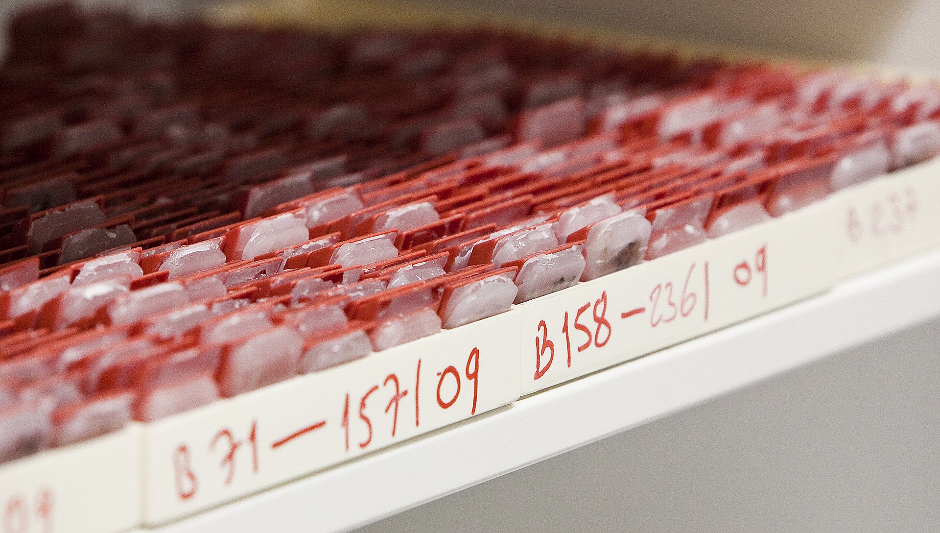
The new technique has been developed in conjunction with the National Genome Research Network (NGFN-Transfer). During the process, the researchers succeeded in tracking the HER2 signal pathway at protein level. For the first time, they were able to extract intact proteins from formalin-fixed, paraffin-embedded tissue samples. “These ‘FFPE’ tissue samples are used as standard in all hospitals,” explains Prof. Becker. “However, up to now it has been very difficult to extract proteins from them.” In order to detect the active HER2 phosphoproteins, the researchers used a combination of biopsies and an automated analysis method known as protein arrays.The research team compared their results with protein analyses of frozen tumor tissue. Unlike FFPE tissue samples, proteins are relatively easy to detect in frozen, untreated biopsies. The activated HER2 molecule was also found in many frozen tumor samples that were previously classified as HER2-negative. “Fresh or frozen tissue samples are rarely found in hospitals,” explains Prof. Becker. “In the future, pathologists will be able to use FFPE tissue, which is routinely prepared during every biopsy, to perform more accurate testing for HER2 using our method. This will give breast cancer patients a better chance of receiving successful treatment.” The methodology is also suitable for other biomarkers, in particular in tissue samples of other types of tumor.
Publication:
Molecular analysis of HER2 signaling in human breast cancer by functional protein pathway activation mapping; Wulfkuhle JD, Berg D, Wolff C, Langer R, Tran K, Illi J, Espina V, Pierobon M, Deng J, Demichele A, Walch A, Bronger H, Becker I, Waldhor C, Hofler H, Esserman LJ, Liotta LA, Becker KF, Petricoin EF 3rd, Clin Cancer Res. (2012); doi:10.1158/1078-0432.CCR-12-0452
Contact Technische Universität München:
Prof. Dr. Karl-Friedrich Becker
Technische Universität München
Institut für Pathologie
Trogerstr. 18
81675 München
Phone: +49 (0) 89 4140 4591
E-mail: kf.becker@lrz.tum.de
Internet: www.path.med.tum.de/
Contact NGFN:
Dr. Cornelia Depner
NGFN Geschäftsstelle
c/o Deutsches Krebsforschungszentrum, V025
Im Neuenheimer Feld 280
69120 Heidelberg
Phone: +49 6221 42-4742
Fax: +49 6221 42-4651
E-mail: c.depner@dkfz.de
Internet: www.ngfn.de